
Onion root tip mitosis labs are common in biology, offering a visual study of cell division.
Resources like Rice University’s wiki and Chegg provide answer keys and guides for analyzing onion cells undergoing mitosis, often in PDF format.
What is Mitosis?
Mitosis is a fundamental process where a single cell divides into two identical daughter cells, crucial for growth and repair in organisms. This process involves several distinct stages – prophase, metaphase, anaphase, and telophase – each characterized by specific chromosomal behaviors.
Understanding mitosis is key to interpreting onion root tip labs, as the actively dividing cells in the root tip provide a clear view of these stages. Many online resources, including those found on Studocu and through university wikis, offer answer keys and detailed explanations to aid in identifying each phase. These PDF guides often include diagrams and tallies to help students accurately assess the proportion of cells in each stage of mitosis.
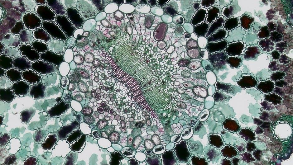
Why Use Onion Root Tips?
Onion root tips are ideal for studying mitosis because they contain a high concentration of rapidly dividing cells, specifically in the meristematic zone. This area facilitates easy observation of the different stages of cell division under a microscope. Prepared slides of onion root tips are commonly used in introductory biology labs, offering a readily available and clear visual representation of mitosis.
Resources like lab manuals and online platforms (Chegg, Studocu) provide answer keys and guides specifically tailored to onion root tip observations, often in PDF format. These materials help students accurately identify and count cells in each mitotic phase, aiding in understanding the duration and frequency of each stage.

The Mitosis Lab: A Step-by-Step Guide
Mitosis labs typically involve preparing onion root tip slides, microscope observation, and data collection. Answer keys, often in PDFs, aid in analysis.
Preparing the Onion Root Tip Slide
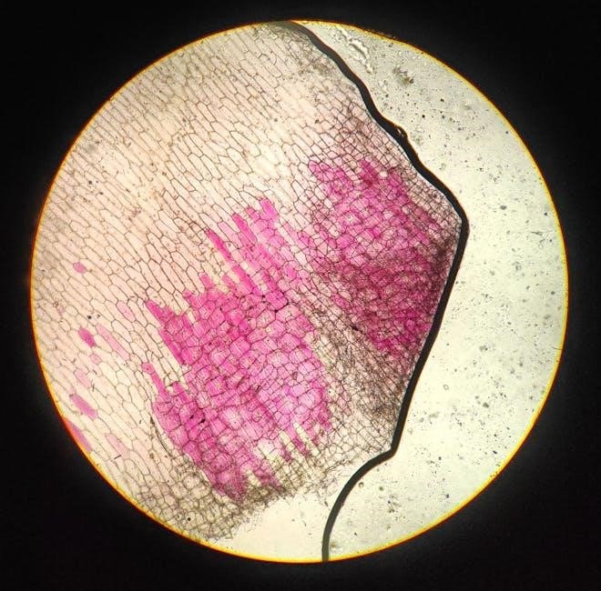
Creating a quality slide is crucial for observing mitosis. Begin by obtaining a prepared slide featuring onion root tips – typically three are present. Carefully examine the slide, holding it to the light to clearly identify the pointed ends of the root sections. These areas are prime locations for actively dividing cells, making them ideal for study.
If preparing a wet mount, gently peel a small section of the onion root tip. Place it on a clean microscope slide, adding a drop of water. Cover with a coverslip, avoiding air bubbles. Resources, including answer keys available in PDF format, often detail this process. Proper slide preparation enhances visibility and accurate identification of mitotic stages.
Microscope Setup and Observation
Begin with the lowest power objective (typically 4x or 10x) to locate the onion root tip on the slide. Gradually increase magnification, focusing carefully to bring cells into clear view. Look for the actively dividing cells within the bracketed area of the root tip, where mitosis is most prevalent.
Utilize proper lighting for optimal observation. Answer keys and lab manuals (often available as PDF downloads) emphasize the importance of adjusting both magnification and illumination. As you observe, systematically scan the slide, noting cells exhibiting different mitotic stages. Careful observation, combined with reference materials, is key to accurate identification and analysis.
Stages of Mitosis in Onion Cells
Mitosis progresses through distinct phases – interphase, prophase, metaphase, anaphase, and telophase – visible in onion root tips. PDF answer keys aid in stage identification.
Interphase: The Preparation Stage
Interphase, though not a stage of mitosis itself, is crucial preparation for cell division. During this extended period, the cell grows, replicates its DNA, and synthesizes proteins necessary for subsequent phases. Observing onion root tip cells, interphase appears as cells with a clearly defined nucleus and nucleolus, but without visible chromosomes.
Students often spend significant time identifying cells in interphase due to its prevalence – it constitutes the majority of cells in a meristematic region like the onion root tip. Answer keys in PDF format, available from resources like Rice University and Chegg, emphasize recognizing the intact nuclear envelope as a key characteristic. Correctly identifying interphase is foundational for understanding the entire mitotic process.
Prophase: Chromosomes Condense
Prophase marks the beginning of visible mitosis. During this stage, the duplicated chromosomes condense, becoming shorter and thicker, and are visible as distinct structures within the nucleus of onion root tip cells. The nuclear envelope begins to break down, and the spindle fibers start to form.
Answer keys, often found as PDF documents from sources like Rice University and Chegg, highlight the appearance of condensed chromosomes as the defining feature of prophase. Students should look for clearly defined, thread-like structures. Correct identification relies on differentiating these condensed chromosomes from the less distinct chromatin seen in interphase. Understanding prophase is essential for tracking chromosome behavior throughout mitosis.
Metaphase: Chromosomes Align
Metaphase is characterized by the alignment of duplicated chromosomes along the metaphase plate – an imaginary line in the middle of the cell. Spindle fibers, originating from the poles of the cell, attach to the centromeres of each chromosome. This precise alignment ensures equal distribution of genetic material to daughter cells during cell division.
Onion cell mitosis answer keys, frequently available as PDFs, emphasize the importance of observing this alignment. Resources like those from Rice University and Chegg detail that correctly identifying metaphase involves noting the organized arrangement of chromosomes. Students should distinguish metaphase from anaphase by confirming that sister chromatids remain attached during this stage.
Anaphase: Sister Chromatids Separate
Anaphase marks the crucial separation of sister chromatids, now considered individual chromosomes, pulled towards opposite poles of the cell by shortening spindle fibers. This ensures each daughter cell receives a complete and identical set of chromosomes. Observing this separation is key to identifying anaphase correctly.
Onion cell mitosis answer keys, often found in PDF format, highlight distinguishing anaphase from metaphase. Resources from sites like Chegg and Rice University emphasize that, unlike metaphase, chromatids are no longer aligned at the center but are visibly moving apart. Correct identification, as detailed in lab guides, is vital for accurate data collection and analysis.
Telophase: Two New Nuclei Form
Telophase is the final stage of nuclear division, characterized by the formation of two distinct nuclei within the cell. Chromosomes arrive at the poles and begin to decondense, becoming less visible. The nuclear envelope reforms around each chromosome set, and the spindle fibers disassemble. This prepares the cell for cytokinesis, the division of the cytoplasm.
Onion cell mitosis answer keys, frequently available as PDF documents, emphasize recognizing these features in telophase. Resources like those found on Studocu and Rice University’s wiki detail how to differentiate telophase from earlier stages. Correctly identifying telophase is crucial for accurate tallying and understanding the complete mitotic process.
Identifying Mitotic Stages
Mitotic stages are identified using prepared slides of onion root tips, aided by answer keys (often PDFs) and tally charts for accurate observation.
Using a Tally Chart for Data Collection
Tally charts are essential tools when analyzing onion root tip mitosis, providing a structured method for recording observations. Students systematically count cells in each mitotic stage – Interphase, Prophase, Metaphase, Anaphase, and Telophase – and record these counts using tally marks.
These charts, often found within lab manuals or answer key PDFs, facilitate quantitative analysis. A key (e.g., “I = Interphase”, “P = Prophase”) is crucial for clarity. Accurate tallying allows for calculating the percentage of cells in each stage, revealing the duration of each phase. Resources like those from Studocu demonstrate this process, emphasizing careful observation and precise recording for meaningful data interpretation.
Key Features to Look For in Each Stage
Identifying mitotic stages in onion cells requires recognizing distinct characteristics. Interphase shows a clear nucleus and no visible chromosomes. Prophase displays condensing chromosomes. Metaphase features chromosomes aligned at the cell’s center. Anaphase is marked by separating sister chromatids moving to opposite poles. Finally, Telophase exhibits two forming nuclei.
Answer key PDFs and resources like those from Rice University often highlight these features. Observing the root tip under a microscope, students should focus on chromosome behavior and nuclear envelope changes. Careful attention to these details, guided by lab manuals and available resources, ensures accurate stage identification and successful data analysis.

Analyzing Mitosis Data
Mitosis rate calculations, often found in onion cell mitosis lab answer key PDFs, involve tallying cells in each stage and determining the percentage in mitosis.
Calculating Mitosis Rate
Calculating the mitosis rate is a core component of the onion root tip lab, and detailed instructions are frequently available within answer key PDF resources. This rate represents the proportion of cells actively undergoing division within the observed field of view. Typically, you’ll begin by totaling the number of cells identified in each stage of mitosis – interphase, prophase, metaphase, anaphase, and telophase.
Then, sum the cells in all mitotic stages (prophase through telophase). Divide this sum by the total number of cells counted (including interphase cells). Finally, multiply the result by 100 to express the mitosis rate as a percentage. Answer keys often provide example calculations and expected ranges for a healthy onion root tip, aiding in result validation.
Interpreting the Results
Interpreting mitosis results from the onion root tip lab involves understanding what the calculated mitosis rate signifies. A higher percentage of cells in mitotic phases suggests rapid growth and active cell division, typical of the root tip’s function. Answer key PDF documents often provide benchmarks for comparison, indicating expected ranges for healthy onion roots.
Deviations from these norms can indicate experimental errors or external factors affecting cell division. Analyzing the distribution of cells across different stages can also reveal insights into the cell cycle’s progression. Resources like those from Rice University emphasize correlating observations with the onion’s growth and development, solidifying the link between mitosis and plant biology.

Common Challenges and Troubleshooting
Identifying dividing cells and distinguishing between mitosis stages can be difficult. Answer key PDF resources and careful observation aid accurate stage identification.
Difficulty Locating Dividing Cells
Locating cells actively undergoing mitosis within the onion root tip requires focused observation. Students often struggle to find areas with visible division, as not all cells are simultaneously dividing. The actively dividing cells are primarily found in the region just behind the root tip’s growing point, as highlighted in lab manuals and online resources.
Prepared slides, while convenient, can sometimes lack optimal staining or sectioning, making identification harder. Utilizing brightfield microscopy and adjusting the light intensity are crucial. Answer key PDF guides often include images pinpointing the meristematic zone where mitosis is most prevalent. Careful scanning of the slide, coupled with referencing these visual aids, significantly improves success in finding dividing cells for accurate analysis.
Distinguishing Between Stages
Differentiating between the stages of mitosis – prophase, metaphase, anaphase, and telophase – demands careful observation of chromosome behavior. Students frequently confuse early prophase with interphase, or metaphase with anaphase.
Key indicators include chromosome condensation (prophase), alignment at the metaphase plate (metaphase), sister chromatid separation (anaphase), and the formation of new nuclei (telophase). Onion cell mitosis answer key PDF resources, like those from Studocu and Rice University, provide detailed diagrams and descriptions of each stage. Comparing observed cells to these visual references is vital. Mastering these distinctions is crucial for accurate data collection and interpreting the mitosis process.
Answer Key Resources & PDF Availability
Onion root tip mitosis labs often have accompanying answer keys, frequently available as PDF documents online. Resources like Chegg and university wikis provide support.
Finding Reliable Answer Keys Online
Locating accurate answer keys for onion root tip mitosis labs requires careful source evaluation. Websites like Chegg offer solutions, but verifying their accuracy is crucial. University campus wikis, such as Rice University’s, often provide student-created resources, potentially offering detailed explanations and PDF versions of answer keys.
However, remember that student-generated content may contain errors. Always cross-reference information with your lab manual and instructor’s guidance. Searching specifically for “onion cell mitosis answer key PDF” can yield results, but prioritize academic sources. Be wary of sites offering complete answers without explanation, as understanding the process is more important than simply obtaining correct results. Look for resources that explain why an answer is correct, not just what the answer is.
Understanding Lab Report Expectations
When completing an onion root tip mitosis lab report, simply copying an answer key PDF is insufficient. Instructors assess your understanding of the mitotic stages, not just your ability to find correct answers. Reports typically require detailed observations, accurate stage identification, and a correctly calculated mitosis rate.
Expectations include clear diagrams illustrating each stage – Interphase, Prophase, Metaphase, Anaphase, and Telophase – with labeled key features. Your tally chart data must be presented logically, and your interpretation should explain the significance of the observed mitosis rate. Avoid plagiarism; use answer keys as a study tool to verify your work, but always express your understanding in your own words.

The Role of Mitosis in Plant Growth
Mitosis drives onion development and root elongation through continuous cell division. Studying onion root tip mitosis, aided by available answer key PDFs, reveals this crucial growth process.
Cell Division and Root Elongation
Mitosis is fundamentally linked to root elongation in onions, as new cells generated through this process contribute directly to increased root length. Observing onion root tip cells under a microscope allows students to visualize the stages of cell division and understand how this process fuels plant growth.
The actively dividing cells are concentrated in the root tip’s meristematic zone, making it an ideal location for studying mitosis. Resources like lab manuals and online platforms, including those offering onion cell mitosis answer key PDFs, provide guidance for identifying and counting cells in each stage of the mitotic cycle.
Understanding the proportion of cells in each phase—interphase, prophase, metaphase, anaphase, and telophase—offers insights into the rate of cell division and, consequently, the rate of root growth. This connection highlights the vital role of mitosis in plant development.
Importance of Mitosis for Onion Development
Mitosis is absolutely crucial for the growth and development of onions, enabling the formation of new cells for bulb expansion, leaf production, and root systems. The consistent cell division within the onion’s meristems, particularly in the root tips, directly impacts the plant’s overall size and yield.
Labs focusing on onion root tip mitosis, often accompanied by answer key PDFs for student support, demonstrate how this process ensures genetic continuity. Each new cell receives an identical set of chromosomes, maintaining the plant’s characteristics.
Resources from institutions like Rice University and platforms like Chegg emphasize the importance of accurately identifying mitotic stages. This understanding is key to appreciating how mitosis drives the onion’s life cycle, from seedling to mature plant.